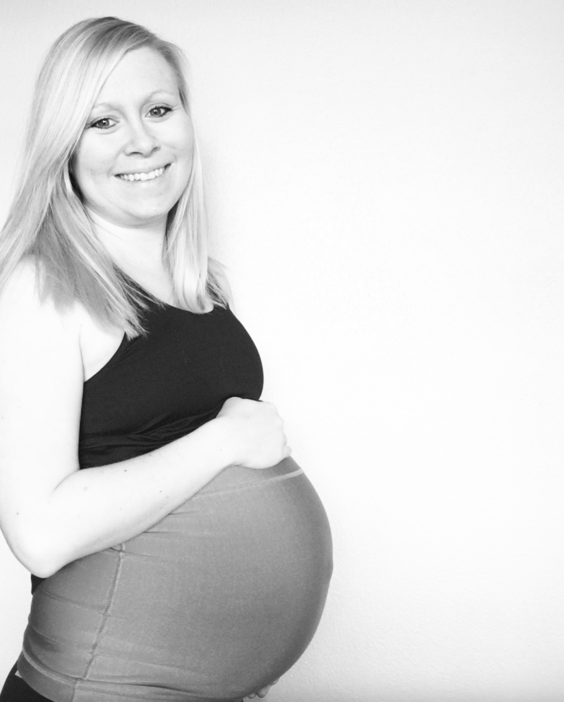
Unlike a lot of pregnant momma's, we found out super late in the game that Lillie had Agenesis of the Corpus Callosum. Many families find out much earlier, during the 20 weeks anatomy scan, or even post-birth. But for us, it was at 33 weeks.
I want to say that our timing was perfect for us. While it was a whirlwind, I felt that we were able to celebrate that we were pregnant, celebrate that we were having our first girl, and primarily wobble through most of my pregnancy without worry. The worry started instantly when we heard the first iterations of our diagnosis (and there were plenty ups and downs).
Also, it was enough time for us to change our birth plan, meet with the specialists, and adjust even where we were giving birth to support our new information of Lillie having ACC.
Our Diagnosis Story
We found enlarged ventricles at 33 weeks, which instantly turned into a whirlwind of an experience. I often think about that day. A full-body anatomy scan that late in the game wasn't in the plans; however, we had experienced a fairly traumatic birth with our older son that lead to a 26 hour labor and vacuum-assisted vaginal delivery — a soft cup with a handle and a vacuum pump literally suction cupping my son out of me, who was stuck. A vacuum extraction poses a risk of injury for both mother and baby, and while I didn't want a cesarean delivery, I also wanted to be mindful of induction dates and whatnot. At 8 lbs. 1 oz. and a 10-day post-due date delivery with my son, the doctors had suspected that his size compared to mine (110 lbs., 5 ft. even) was one of the main factors. I was measuring larger than I should at 33 weeks, and I had asked that day to see how big they thought the baby really was — thus an anatomy scan.
Hydrocephalus, or enlarged ventricles, is the abnormal enlargement of the brain cavities (ventricles) caused by a build-up of cerebrospinal fluid. I was familiar with it before as I had a grandpa who had "water on the brain" and needed a shunt to drain much of the fluid in his later years of life. In adults 60 years of age and older, the more common signs and symptoms of hydrocephalus are loss of bladder control, memory loss, progressive loss of other thinking or reasoning skills, and difficulty walking. Yet I had no idea what it meant for a baby.
We were immediately recommended to our Maternal Fetal Medicine providers and found ourselves in the midst of testings and visits with the specialists at Fetal Care Center at St. Louis Children's Hospital. We were instantly told that no, Lillie did not have hydrocephalus, but more so ventriculomegaly. Ventriculomegaly is a condition in which the ventricles appear larger than normal on a prenatal ultrasound. This can occur when CSF becomes trapped in the spaces, causing them to grow larger. This condition occurs in approximately one in 1,000 infants. Typically, ventriculomegaly only requires treatment if it causes hydrocephalus, so the diagnosis of hydrocephalus itself was a bit premature. Additionally through a Level 2 ultrasound, the MFM office confirmed we would need additional testing to see if Lillie's corpus callosum was present. A Level 2 ultrasound is a comprehensive, detailed evaluation of fetal anatomy and development. It is a much more in-depth evaluation of the fetus than a standard or Level 1 ultrasound that you would find in a standard prenatal screening.
By these appointments, we were nearing 35 weeks and could not conduct any type of extensive genetic testing without it risking preterm labor. We had opted out of the testing earlier in our pregnancy with both kids, and was told we would need to wait for the doctors to take blood samples from the umbilical cord before they could conduct much further testing at all. Even amniocentesis testing was risky at this late in our pregnancy.
They threw out diagnoses like Down syndrome, Dandy–Walker syndrome, Trisomy 9, and cystic fibrosis, but wouldn't confirm or rule out anything. It was a really hard time of absorbing a lot of scary diagnoses but only being able to "wait and see".

Our MRI Scan
After a round of specialists and a ton of more visits that provided little to no hope, we had scheduled our prenatal MRI to confirm Lillie's missing corpus callosum. At the time, we were told that to determine if her corpus callosum was missing, shortened, thinned, or thickened would tell a lot about her life once she was born. While there isn't much data on any of the various cases, from our knowledge, being born without it completely was the best case scenario.
The prenatal MRI was hard. Laying still on my back for what felt like forever that pregnant was sickening. I felt the instant desire to throw up with the amount of acid reflux in my system. It was painful, uncomfortable, and I could not breathe. It was loud, nerve-racking, and down right no fun. And then we had to return a full 24 hours later to have the results read to us.
As we met with our neurologist the following day, our doctor explained that Lillie had complete agenesis of the corpus callosum, meaning her corpus callosum was complete absent. It is also seen at times as C-ACC or CACC for 'complete'. In addition, they confirmed that Lillie was also missing her cavum septum pellucidum (CSP).
By itself, we were told, absence of the septum pellucidum (CSP) was not life-threatening, or much of a concern. When it is missing, symptoms may include learning difficulties, behavioral changes, seizures, and changes in vision. Absence of the septum pellucidum, however, is not typically seen as an isolated finding. And for us, the absence of the septum pellucidum is associated with other conditions, like her missing corpus callosum, added another diagnosis to our list — septo-optic dysplasia.
We were not given much information about Lillie's septo-optic dysplasia diagnosis at that time. The doctors were really focused on ensuring we were prepared for delivery and our birth plan going into the upcoming weeks.
Our Change in Birth Plan
We had the most convenient hospital near our home. It was new, great doctors, and allowed for private birthing rooms that transitioned into your recovery room post-birth. We could "move in" for a few days and never need to be transitioned to a new room, and was only about a ten minute drive home. That all changed once we received Lillie's diagnosis.
Once we found out about Lillie's ACC, we met with many specialists including a lead neurologist and his team, an ophthalmologist, genetic's counselor, endocrinologist, and many various MFM doctors and nurses. They talked about the importance of being near a NICU "just in case" something happened. While they did tell us that Lillie's birth could be easy and non-traumatic, if anything was to happen with her breathing or eating abilities, she would be transferred away from us immediately.
Thankfully, we were near enough to make the decision that was right for us. St. Louis Children's Hospital had just opened up their new Parkview Tower with their labor and delivery rooms. It also directly connected to the their newly expanded Newborn Intensive Care Unit (NICU) in St. Louis Children’s Hospital, making it easy for both me to visit our baby. Simply put, I did not need to be discharged from my labor and delivery to see my NICU-potential baby and all of her doctors could take any testing they needed to prior to Lillie and I being discharged from her birth. This specifically was huge for us, as the alternative, even with a healthy delivery, would have put us carrying a 3 to 4 day old newborn to the Children's Hospital for post-birth MRI testing and more. With the switch we were able to have it all done while we were healing and bonding together.


2 comments